The ears are paired sensory organs comprising the auditory system, involved in the detection of sound, and the vestibular system, involved with maintaining body balance/ equilibrium. The ear divides anatomically and functionally into three regions: the external ear, the middle ear, and the inner ear. All three regions are involved in hearing. Only the inner ear functions in the vestibular system.
Anatomy of the Ear
The external ear (or pinna, the part you can see) serves to protect the tympanic membrane (eardrum), as well to collect and direct sound waves through the ear canal to the eardrum. About 1¼ inches long, the canal contains modified sweat glands that secrete cerumen, or earwax. Too much cerumen can block sound transmission.

The middle ear, separated from the external ear by the eardrum, is an air-filled cavity (tympanic cavity) carved out of the temporal bone. It connects to the throat/nasopharynx via the Eustachian tube. This ear-throat connection makes the ear susceptible to infection (otitis media). The eustachian tube functions to equalize air pressure on both sides of the eardrum. Normally the walls of the tube are collapsed. Swallowing and chewing actions open the tube to allow air in or out, as needed for equalization. Equalizing air pressure ensures that the eardrum vibrates maximally when struck by sound waves.
Adjoining the eardrum are three linked, movable bones called "ossicles," which convert the sound waves striking the eardrum into mechanical vibrations. The smallest bones in the human body, the ossicles are named for their shape. The hammer (malleus) joins the inside of the eardrum. The anvil (incus), the middle bone, connects to the hammer and to the stirrup (stapes). The base of the stirrup, the footplate, fills the oval window which leads to the inner ear.
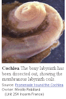
The inner ear consists of a maze of fluid-filled tubes, running through the temporal bone of the skull. The bony tubes, the bony labyrinth, are filled with a fluid called perilymph. Within this bony labyrinth is a second series of delicate cellular tubes, called the membranous labyrinth, filled with the fluid called endolymph. This membranous labyrinth contains the actual hearing cells, the hair cells of the organ of Corti.
There are three major sections of the bony labyrinth:
1. The front portion is the snail-shaped cochlea, which functions in hearing.
2. The rear part, the semicircular canals, helps maintain balance.
3. Interconnecting the cochlea and the semicircular canals is the vestibule, containing the sense organs responsible for balance, the utricle and saccule.
The external ear (or pinna, the part you can see) serves to protect the tympanic membrane (eardrum), as well to collect and direct sound waves through the ear canal to the eardrum. About 1¼ inches long, the canal contains modified sweat glands that secrete cerumen, or earwax. Too much cerumen can block sound transmission.

The middle ear, separated from the external ear by the eardrum, is an air-filled cavity (tympanic cavity) carved out of the temporal bone. It connects to the throat/nasopharynx via the Eustachian tube. This ear-throat connection makes the ear susceptible to infection (otitis media). The eustachian tube functions to equalize air pressure on both sides of the eardrum. Normally the walls of the tube are collapsed. Swallowing and chewing actions open the tube to allow air in or out, as needed for equalization. Equalizing air pressure ensures that the eardrum vibrates maximally when struck by sound waves.
Adjoining the eardrum are three linked, movable bones called "ossicles," which convert the sound waves striking the eardrum into mechanical vibrations. The smallest bones in the human body, the ossicles are named for their shape. The hammer (malleus) joins the inside of the eardrum. The anvil (incus), the middle bone, connects to the hammer and to the stirrup (stapes). The base of the stirrup, the footplate, fills the oval window which leads to the inner ear.
The inner ear consists of a maze of fluid-filled tubes, running through the temporal bone of the skull. The bony tubes, the bony labyrinth, are filled with a fluid called perilymph. Within this bony labyrinth is a second series of delicate cellular tubes, called the membranous labyrinth, filled with the fluid called endolymph. This membranous labyrinth contains the actual hearing cells, the hair cells of the organ of Corti.
There are three major sections of the bony labyrinth:
1. The front portion is the snail-shaped cochlea, which functions in hearing.
2. The rear part, the semicircular canals, helps maintain balance.
3. Interconnecting the cochlea and the semicircular canals is the vestibule, containing the sense organs responsible for balance, the utricle and saccule.
The inner ear has two membrane-covered outlets into the air-filled middle ear - the oval window and the round window. The oval window sits immediately behind the stapes, the third middle ear bone, and begins vibrating when "struck" by the stapes. This sets the fluid of the inner ear sloshing back and forth. The round window serves as a pressure valve, bulging outward as fluid pressure rises in the inner ear. Nerve impulses generated in the inner ear travel along the vestibulocochlear nerve (cranial nerve VIII), which leads to the brain. This is actually two nerves, somewhat joined together, the cochlear nerve for hearing and the vestibular nerve for equilibrium.
How We Hear - The Auditory System

All sounds (music, voice, a mouse-click, etc.) send out vibrations, or sound waves. Sound waves do not travel in a vacuum, but rather require a medium for sound transmission, e.g. air or fluid. What actually travels are alternating successions of increased pressure in the medium, followed by decreased pressure. These vibrations occur at various frequencies, not all of which the human ear can hear. Only those frequencies ranging from 20 to 20,000 Hz (Hz = hertz = cycles/sec) can be perceived.
In hearing, air-borne sound waves funnel down through the ear canal and strike the eardrum, causing it to vibrate. The vibrations are passed to the small bones of the middle ear (ossicles), which form a system of interlinked mechanical levers: First, vibrations pass to the malleus (hammer), which pushes the incus (anvil), which pushes the stapes (stirrup). The base of the stapes rocks in and out against the oval window - this is the entrance for the vibrations. The stapes agitates the perilymph of the bony labyrinth. At this point, the vibrations become fluid-borne. The perilymph, in turn, transmits the vibrations to the endolymph of the membranous labyrinth and, thence, to the hair cells of the organ of Corti. It is the movement of these hair cells which convert the vibrations into nerve impulses. The round window dissipates the pressure generated by the fluid vibrations, thus serves as the release valve: It can push out or expand as needed. The nerve impulses travel over the cochlear nerve to the auditory cortex of the brain, which interprets the impulses as sound.
How We Balance - The Vestibular System
The semicircular canals and vestibule function to sense movement (acceleration and deceleration) and static position. The three semicircular canals lie perpendicular to each other, one to sense movement in each of the 3 spatial planes. At the base of the canals are movement hair cells, collectively called the crista ampullaris. Depending on the plane of movement, the endolymph flowing within the semicircular canals stimulates the appropriate movement hair cells. Static head position is sensed by the vestibule, specifically, its utricle and saccule, which contain the position hair cells. Different head positions produce different gravity effects on these hair cells. Small calcium carbonate particles (otoliths) are the ultimate stimulants for the position hair cells.
The hair cells for both position and movement create nerve impulses. These impulses travel over the vestibular nerve to synapse in the brain stem, cerebellum, and spinal cord. No definite connections to the cerebral cortex exist. Instead, the impulses produce reflex actions to produce the corrective response. For example, a sudden loss of balance creates endolymph movement in the semicircular canals that triggers leg or arm reflex movements to restore balance.
Adapted from: PATTS
